Breakthrough in Bioprinting
A new technique raises hope for repairing damaged tissue.
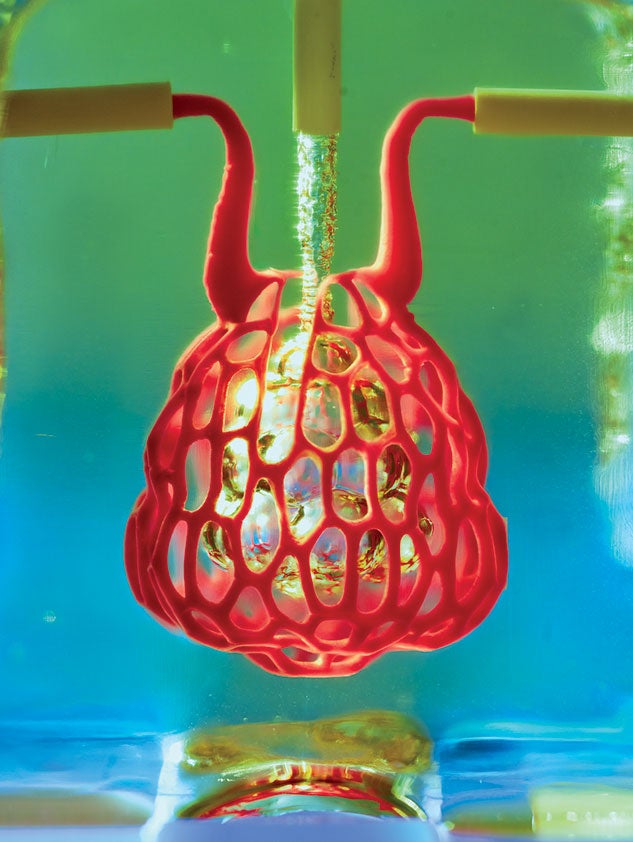
In the lab of Rice bioengineer Jordan Miller, a tiny teardrop-shaped air sac expands and contracts in a steady rhythm. Blood flows through a shroud of vessels surrounding it. Though the entwined realms of blood and air never meet, life-giving oxygen moves from one to the other. This model represents a big leap in 3D printing technology for living tissue — and promises new treatments for diseases of the liver, heart, lungs and more.
Bioprinting living tissue could one day help in repairing or replacing damaged organs. The innovative technique for creating entangled
vascular networks was created by Miller and bioengineer Kelly Stevens of the University of Washington (UW), along with 15 collaborators from Rice, UW, Duke University, Rowan University and Nervous System, a design firm in Somerville, Mass.
“One of the biggest roadblocks to generating functional tissue replacements has been our inability to print the complex vasculature that can supply nutrients to densely populated tissues,” said Miller, an assistant professor of bioengineering. “Our organs actually contain independent vascular networks — like the airways and blood vessels of the lung or the bile ducts and blood vessels in the liver. These interpenetrating networks are physically and biochemically entangled, and the architecture itself is intimately related to tissue function.”
“Tissue engineering has struggled with multivascularization for a generation,” Stevens said. “With this work we can now better ask, ‘If we can print tissues that look and now even breathe more like the healthy tissues in our bodies, will they also then functionally behave more like those tissues?’ This is an important question, because how well a bioprinted tissue functions will affect how successful it will be as a therapy.” The research was featured on the cover of the May 3 issue of Science.
The team created a new open-source bioprinting technology dubbed the “stereolithography apparatus for tissue engineering,” or SLATE. The system uses additive manufacturing to make soft hydrogels one layer at a time. Layers are printed from a liquid pre-hydrogel solution that becomes a solid when exposed to blue light.
The key insight by Miller and Bagrat Grigoryan, a Rice graduate student and lead co-author of the research study, was the addition of food dyes that absorb blue light. Tests of the lung-mimicking structure showed that the tissues were sturdy enough to avoid bursting during blood flow and pulsatile “breathing,” a rhythmic intake and outflow of air that simulated the pressures and frequencies of human breathing.
Miller, a long-standing champion of open-source 3D printing, said all source data from the experiments in the published Science study are freely available. “We are only at the beginning of our exploration of the architectures found in the human body,” he said. “We still have so much more to learn.”
The work was supported by the Robert J. Kleberg, Jr. and Helen C.
Kleberg Foundation, the John H. Tietze Foundation, the National Science Foundation, the National Institutes of Health and the Gulf Coast Consortia.
— Jade Boyd